



Multi-spot OCT tracks cornea deformations
ICTER project develops biomechanical mapping method as clinical diagnosis tool.
29 July 2026
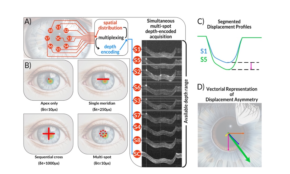
Comparison of conventional OCT approaches with a new multi-spot method that simultaneously measures air-puff-induced corneal deformation at nine locations. This enables assessment of corneal asymmetry, relevant particularly to keratoconus. Credit: Biomedical Optics Express.
Researchers at the International Centre for Translational Eye Research (ICTER), part of Poland's Institute of Physical Sciences, have developed a prototype OCT imaging system able to track mechanical responses of the eye's cornea at nine locations simultaneously.
The technology marks an important step toward more accurate, non-contact assessment of corneal biomechanics, said the project, and could improve early detection and monitoring of disorders such as keratoconus in which the cornea becomes thinned and deformed.
Assessing the physical and biomechanical properties of structures in the eye has become increasingly important in modern ophthalmology, revealing changes that can appear before visible structural abnormalities develop. However, accurately measuring these properties in the cornea remains technically challenging.
Described in Biomedical Optics Express, the ICTER method exploits developments in swept-source OCT platforms, where VCSEL-based lasers have brought about significant increases in the coherence length available from an OCT approach. ICTER combined this with space-division multiplexing, a technique from datacomms here repurposed for optical metrology allowing multiple spatial locations to be examined at the same time.
Rather than illuminating the cornea with a single probing beam, the ICTER system strikes the tissue with several beams simultaneously at different measurement locations. The signals are separated using depth encoding, so the platform need not acquire data from the locations sequentially but instead record data from multiple points within a single OCT acquisition.
The project used air-puff stimulation, as employed by ophthalmologists during the measurement of intraocular pressure, to apply physical force to the cornea, and monitored the response simultaneously at nine positions: one central point and eight surrounding peripheral locations.
New details of complex cornea dynamics
A spread of locations made it possible not only to determine how much the cornea deforms but also to identify precisely where the tissue responds most strongly, noted the team, and whether that response may indicate local biomechanical weakening. By recording all nine locations simultaneously, the ICTER system eliminates temporal inconsistencies and provides a much more faithful representation of how the cornea responds to external force.
"The corneal deformation we aim to capture to assess its mechanical asymmetry lasts only about 20 milliseconds, and the measurement cannot be repeated," commented ICTER's Karol Karnowski. "This makes the simultaneous acquisition of the corneal response at multiple locations the key feature of our method. Achieving this requires very high temporal resolution, and existing techniques have either lacked this temporal resolution or have been limited in their spatial coverage."
The ICTER results revealed unexpected details of the cornea's biomechanical response to the stimulation, with not one but two clearly distinguishable deformation phases following an air pulse. Because nine locations were recorded simultaneously at high temporal resolution, the researchers were able to observe this phenomenon directly rather than infer it retrospectively from sequential scans, an example of the subtle dynamic phenomena that this approach may reveal.
Having now tested the underlying principle on the cornea, ICTER predicts that the new approach could have applications beyond ophthalmology.
This concept could prove valuable for studying other soft tissues where fast mechanical processes are difficult to capture reliably with existing imaging techniques, commented the team, or in scenarios where conventional sequential scanning may introduce significant temporal errors.
"As the technology continues to mature and undergo clinical validation, it could become a valuable addition to the ophthalmic diagnostic toolbox, enabling clinicians to detect biomechanical abnormalities earlier, monitor disease progression more precisely, and make better-informed treatment decisions," wrote ICTER.
OCT with tunable lens allows whole-eye imaging
January 25 2018
New VCSEL offers simpler, cheaper OCT
June 05 2023

Syntec swings to profit as sales climb
August 19 2026



Hesai goes ‘full-stack’ with extended robotics offering
August 19 2026