


Rockefeller University microscope points to deeper brain imaging
Redesigned three-photon platform gathers data at "unprecedented tissue depth."
04 November 2026
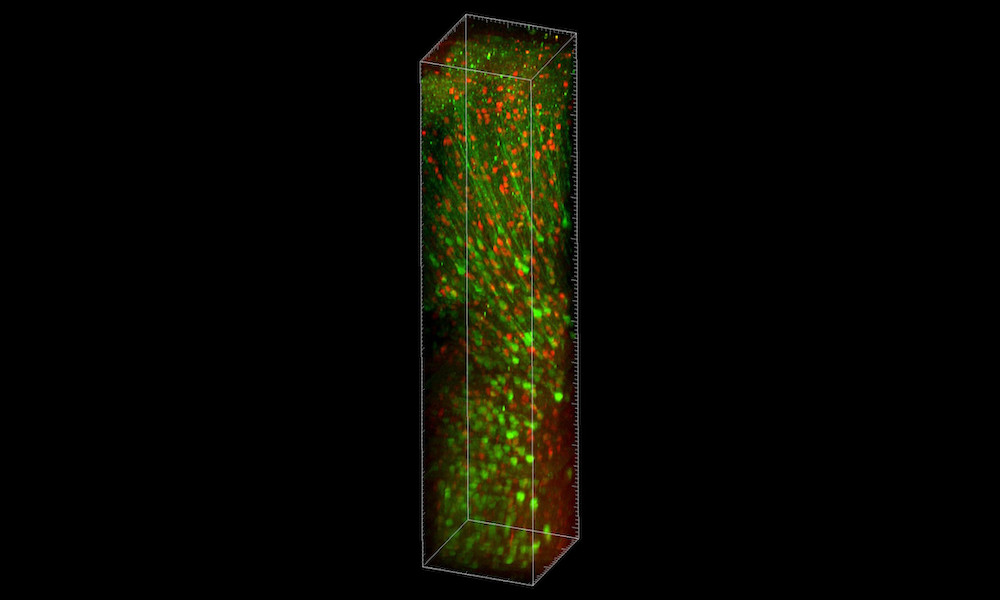
A volumetric rendering of stacked images taken with the Vaziri lab’s new platform TrUDI. Credit: Rockefeller University/Vaziri Lab.
A project at the Rockefeller University Laboratory of Neurotechnology and Biophysics has developed an imaging platform designed to extend the reach of multiphoton microscopy to deeper brain tissues.
One primary goal was to tackle the fundamental hurdle in deep-brain imaging, by which reaching greater depths typically requires more laser power. This can in turn cause overheating and damage living brain tissue.
The Rockefeller team has now "redesigned the entire imaging system from the ground up," and published its findings in Nature Methods.
Its innovative microscopy platform has been christened Three-photon Ultra-Deep Imaging, or TrUDI, and pushes three-photon imaging close to its theoretical limits, noted the team. This has been achieved in part by reengineering the rate and energy of laser pulses, along with how they are delivered and scanned at the sample, all within the safe limits of sample power exposure.
"Often in technology, people stick with a certain laser or parameter for historical reasons, and everyone just accepts those limitations," said Alipasha Vaziri, head of the Laboratory of Neurotechnology and Biophysics.
"We wanted to take a clean slate approach. By not taking anything for granted and asking how a system fully optimized for maximizing the depth reach of three-photon imaging would look like from scratch, we found that what people previously thought to be limiting factors could be overcome."
Three-photon microscopy was designed to extend the ~0.8 millimeter depth reach of the more widely used two-photon techniques. But until now three-photon fluorescence has hit natural limits, relating to the amount of laser power needed to create the simultaneous absorption of three excitation photons upon which the method hinges. As a result the three-photon approach has been falling short of its potential.
Bringing new brain regions within reach
"In any multi-photon excitation scheme, there is a depth limit at which the background and the signal become equal," commented Vaziri. "For three-photon excitation in the mouse brain, theoretically that depth limit should be on the order of 2.3 millimeters. But the maximum depth at which functional recording has been previously achieved was about 1.2 millimeters."
The Rockefeller redesign involved first using computer models to determine the theoretical limits of deep-brain imaging and identify how a microscope would need to be designed to approach them. Guided by those calculations, the project went back to the drawing board and optimized every aspect of three-photon microscopy, most notably by delivering fewer but much more energetic laser pulses while maintaining the same average laser power, and by redesigning how the microscope focuses and collects light.
The team also developed an excitation scheme that identifies neurons and activates the laser only when passing over those cells, reducing laser exposure by up to 80 percent during long sessions.
As a result the TrUDI platform boasts a roughly 250-fold increase in fluorescence photon yield without relying on increased laser power, achieving cellular-resolution recordings of neuronal population activity at tissue depths of up to ~2 millimeters in mouse cortex and hippocampus.
"There are regions of the striatum that are inaccessible, and there are still a number of deep brain nuclei which are much deeper that still require invasive approaches," commented Vaziri. "But our work brings a series of brain regions that have been previously accessible to imaging only through invasive means within reach, without that kind of invasiveness."
Helmet-mounted sensors reveal how brain learns new tasks
October 29 2026

Laser weapons and battery welding aid IPG Photonics sales growth
November 04 2026


Europe-Japan group to industrialize flexible perovskite solar cells
November 03 2026

Marvell set to acquire Lumentum in $115BN all-stock deal
November 03 2026