


SPIE O+P 2026: AI-based image analysis for bio-imaging
Prof. Arrate Muñoz-Barrutia, of Universidad Carlos III de Madrid, discusses how AI is transforming biological data.
26 August 2026
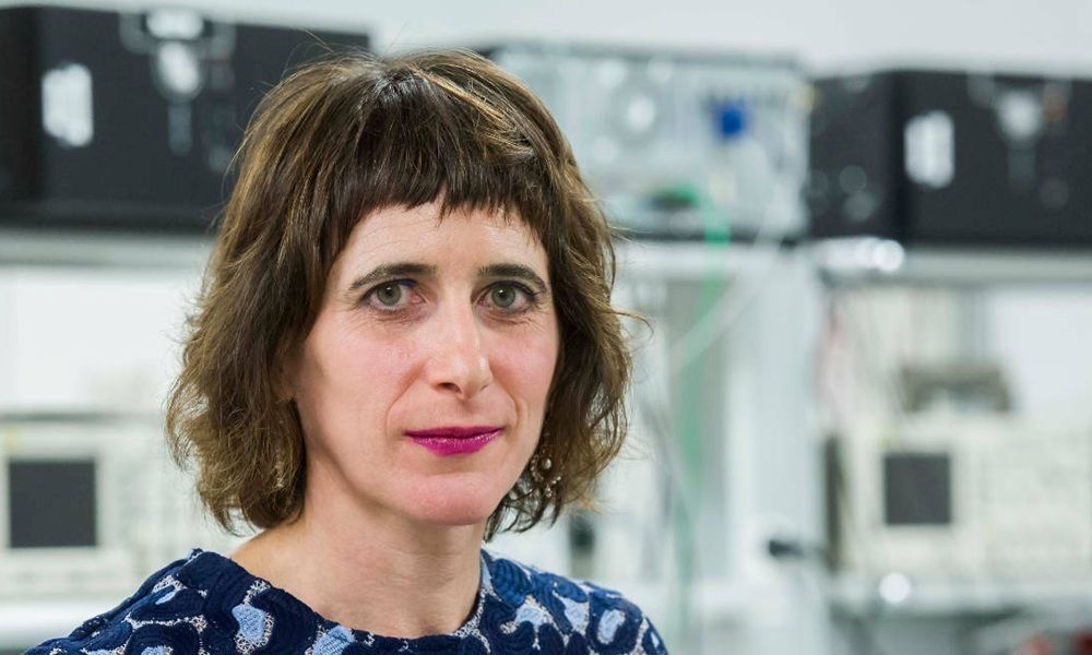
Professor Arrate Muñoz Barrutia of Universidad Carlos III de Madrid, Spain. Photo: SPIE.
Arrate Muñoz Barrutia is Full Professor at
Muñoz-Barrutia will discuss “Artificial intelligence for microscopy: from image analysis to intelligent imaging” on 26 August at SPIE Optics + Photonics as part of the Artificial Intelligence Plenary and will present “Sleep qEEG as a non-invasic biomarker for Alzheimer’s disease” on 27 August.
Ahead of the conference and exhibition, held annually at the San Diego Convention Center, SPIE interviewed Prof. Muñoz-
SPIE: At Optics + Photonics, you will be discussing how interdisciplinary approaches using AI-based image analysis are expanding data-driven discovery in biology and medicine. What is a specific piece of information that you have been able to access using this approach that wasn’t possible prior to this type of image analysis?
Muñoz-Barrutia: For me, this evolution is closely connected to the Cell Tracking Challenge, whose organizing committee I am part of. For many years, we worked on problems such as segmenting and tracking cells in noisy, low-contrast, highly textured microscopy images, but classical algorithms often struggled to generalize across datasets.
A major turning point came in 2015, when U-Net achieved a striking improvement in whole-cell segmentation in phase-contrast images. Since then, I have seen AI move the field from solving long-standing problems more reliably to opening entirely new possibilities.
In our InterpolAI work, for example, deep learning can restore damaged tissue sections and estimate missing information, helping us reconstruct continuous 3D tissue architecture and reveal spatial relationships that could not previously be recovered with confidence.
You will also be presenting a paper you co-authored about using sleep qEEG as a non-invasive biomarker for Alzheimer's disease. Is that one of the areas in which the AI-based image analysis you’re discussing in your plenary has opened new opportunities in medicine?
Sleep qEEG is not image analysis in the strict sense, but it follows the same data-driven approach: using machine learning to detect subtle patterns in complex biomedical data that are difficult to identify visually or with conventional methods.
By analyzing changes in brain activity across different sleep stages, we can identify signatures associated with Alzheimer’s disease before symptoms become pronounced. In the future, this could provide an accessible, non-invasive way to identify people who may benefit from further testing at an earlier stage.
What do you hope people attending your talks at Optics + Photonics will come away discussing or considering?
I hope people will come away thinking about AI not as a stand-alone algorithm, but as part of a complete scientific workflow. Progress depends on bringing together imaging technology, high-quality data, biological and clinical knowledge, robust validation, and tools that researchers can actually use. I would especially like attendees to consider how optics, photonics, and AI can be designed together from the beginning to reveal biological information that neither field could access alone.
Is there something you’re hoping to get out of attending Optics + Photonics?
I am especially looking forward to meeting researchers developing new optical and photonic technologies and exploring how these advances can be combined with AI from the earliest stages of an experiment. I hope to discover new approaches, exchange ideas beyond my immediate field, and build collaborations that connect imaging, computation, biology, and medicine.
For you personally, what is one of your proudest achievements, even if it is outside of your research?
One of my proudest achievements is seeing the people I have mentored grow into confident, independent researchers and begin building careers of their own. UC3M places great value on giving undergraduate students both international experience and early contact with research, and our collaborations with institutions such as Johns Hopkins University have helped make those opportunities very real.
Some students have gone directly from their degree into master’s programs at leading universities, while others have entered highly competitive PhD programs at places such as Johns Hopkins or within the MIT–Harvard ecosystem. Watching them discover what excites them, take ambitious steps, and eventually begin supporting others is one of the most rewarding parts of my career.
Having a strong support network is an important component in research and science. Who are some key players in your support network and how do they support you?
I have been fortunate to learn from many people throughout my career. My PhD supervisor, Professor Michael Unser at EPFL, gave me a rigorous foundation in image processing and taught me the value of technical excellence. Later, Dr. Carlos Ortiz de Solórzano, my postdoctoral supervisor at CIMA–University of Navarra, helped me connect engineering with meaningful biological questions.
More recently, Professor Denis Wirtz at Johns Hopkins University has been a fundamental source of support and inspiration, shaping a way of doing science that is creative, collaborative, and always open to learning. Beyond these mentors, my wider international network of collaborators — across research consortia, joint projects, and the institutions where I have worked or visited — has continually challenged me, encouraged me, and helped me grow.
What do you do in your free time to disconnect from your research?
Physical activity is one of the best ways for me to disconnect. I enjoy hot yoga, Radikal Training, and Pilates Reformer because they require my full attention and help me leave work behind for a while. I also love visiting museums and contemporary art exhibitions. Living in Madrid is a privilege, with places such as the Reina Sofía, the Thyssen-Bornemisza, and the Prado always nearby. Art allows me to look at images in a completely different way — not to measure or analyze them, but simply to enjoy what they make me feel and think.
Lastly, what are some of your takeaways from being part of the SPIE community?
My connection with SPIE goes back to the early stages of my career, when I presented work on mathematical imaging and image processing. What I value most about the SPIE community is the way it brings together fundamental advances in optics and photonics with real applications in biology and medicine. It is also a welcoming environment for exchanging ideas across disciplines, receiving constructive feedback, and supporting young researchers as they take their first steps in the international scientific community.
U.S. Army Research Office awards $19.2 million to Florida team
October 14 2026
ASML looks to add more capacity as chip boom continues
October 14 2026

UK’s Salience Labs adds $125M series B
October 14 2026
Francis Halzen awarded Nobel Prize in Physics 2026
October 12 2026