


Texas A&M sees motion and chemistry together in living tissue
New high-speed movies could help scientists study disease and fast dynamic processes.
14 July 2026
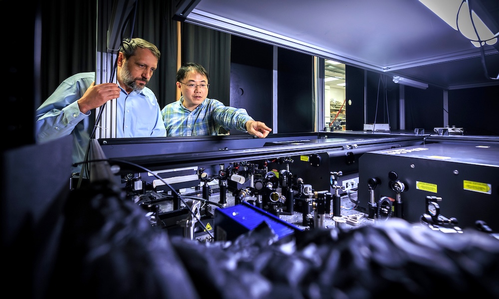
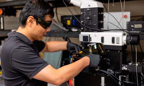
Drs. Alexei Sokolov (left) and Zhenhuan Yi examine the lab’s custom optical system used to generate and measure light for high-speed imaging experiments. Credit: Texas A&M University.
An advanced imaging method developed at Texas A&M University captures chemical processes in living organisms in real time, allowing researchers to "observe biology as it happens."
A range of optical techniques have been applied to the imaging of in vivo dynamics, thanks to the ability of microspectroscopy methods such as Raman and infrared-based techniques to map the chemical distribution of samples based on molecular vibrations without labeling.
However, imaging fast dynamics in living organisms remains challenging, noted the Texas A&M team, and many chemical and biological processes occur too fast to be captured by conventional spectroscopic imaging methods that need scanning or long/multiple exposures.
The project's solution, described in PNAS, is a wide-field infrared microspectroscopy approach to single-shot imaging, where each image is captured with a single pair of laser pulses lasting approximately one picosecond. This minimizes motion blur and allows observation of fast dynamic processes at frame rates up to the laser repetition rate.
"We're able to follow processes that were essentially invisible before," said Alexei Sokolov, associate director of Texas A&M's Institute for Quantum Science and Engineering. "Not just where things are but how they’re evolving, how the underlying chemistry is changing from moment to moment."
The approach is based on a third-order sum-frequency process by which incoming infrared light can be converted to visible signals. Instead of scanning across a sample, the system captures an entire image in a single shot from this resonant emission. Each frame is recorded on the scale of a picosecond, minimizing motion blur.
"At that timescale, things don't have time to move enough to blur the image," Sokolov said. "So what you capture is very close to the system’s natural state."
Tracking how diseases emerge and evolve
In trials the team used its technique to demonstrate single-shot in vivo imaging of live C. elegans worms in water, achieving a spatial resolution of approximately 400 nanometers. Using a kHz laser system allowed video sequences at the same frame rate to be formed from single-shot images, showing the organisms in motion while preserving chemical detail without the distortion typically caused by motion blur.
Crucially, this technique can be applied to water-rich systems and is compatible with aqueous environments, meaning it can image biological samples in vivo with submicron spatial resolution. This opens opportunities for studying fast processes in living systems and complex materials.
It may prove particularly significant as a new means of tracking how diseases emerge and evolve, in systems where timing and chemical interactions are critical. The technique may also support efforts to detect subtle changes in cells earlier or monitor how they respond to therapies, areas where capturing real-time chemical activity has been a longstanding challenge.
The team is now working to expand the method’s capabilities, including how to distinguish more precisely between types of molecules and increasing the sensitivity. The same imaging principle may also be valuable outside biology, noted the Texas A&M team, and can be applied to systems where chemistry changes rapidly in physics and materials science.
"This is really about accessing a different timescale," commented Alexei Sokolov. "Once you can do that, a lot of new questions become possible. This changes what’s observable and gives us a way to study those dynamics directly, instead of inferring them."

Multi-spot OCT tracks cornea deformations
July 29 2026
Gooch & Housego poised for private equity buyout
July 29 2026