


Tufts University observes Herpes virus reshaping neurons
Two-photon and fluorescence lifetime imaging show virus altering brain metabolism before cells are damaged.
26 August 2026
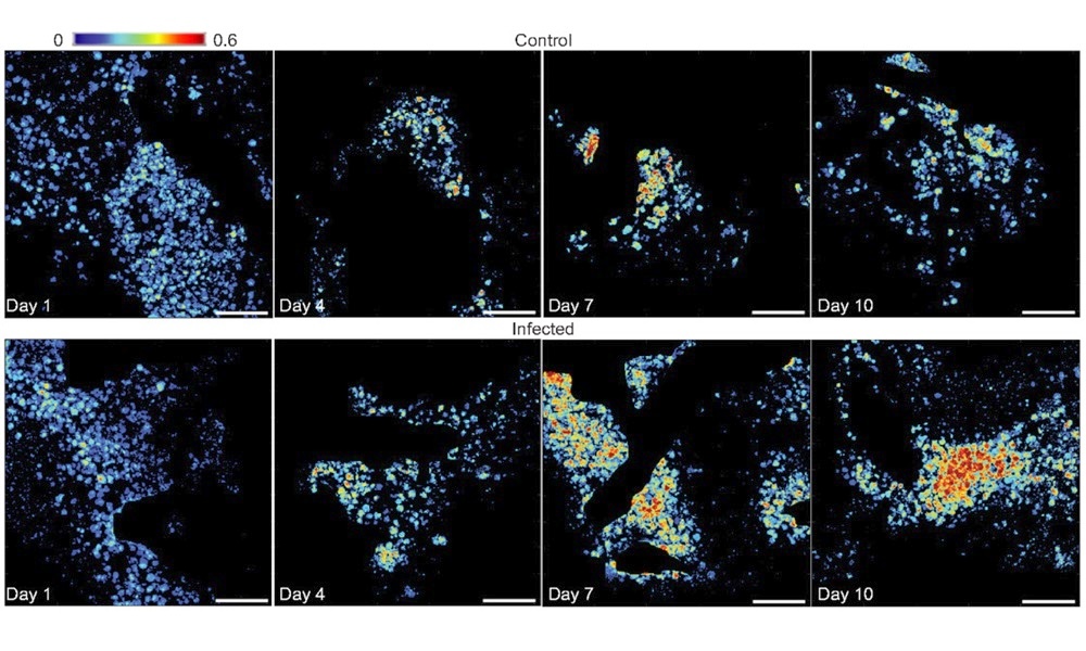
Optical metrics from two-photon excited fluorescence reveal metabolic perturbations during HSV-1 infection. Credit: M. Savvidou/Neurophotonics.
Herpes simplex virus (HSV-1) is known to be a potential factor in neurodegeneration and conditions such as Alzheimer's disease, but the exact mechanisms and metabolic consequences of infection are poorly defined.
A project at Tufts University has now used optical imaging to follow and quantify the ways that HSV-1 induces metabolic reprogramming in human neuronal tissue, and published the results in Neurophotonics.
The team employed two-photon excited fluorescence (TPEF) and fluorescence lifetime image (FLIM) in its study. TPEF allows non-invasive quantitative optical imaging of metabolism with subcellular resolution, by capturing the autofluorescence of two nucleotides known to be key mitochondrial cofactors.
FLIM provides complementary data about metabolic processes, by distinguishing between different molecular states of those cofactors. The technique's sensitivity to environmental parameters including pH, viscosity and temperature gives further data about factors liable to influence cellular metabolism and function.
Tufts researchers grew human neurons in three-dimensional scaffolds that mimic some of the structure of brain tissue, and exposed the tissues to low levels of HSV-1. The optical methods were then used to image the tissues over 10 days.
Since this imaging approach relies on the natural light emitted by molecules involved in energy production, repeated study of the same living tissue could be made over periods of time. This let researchers image the dynamic metabolic response to infection, rather than relying on single end-point measurements.
The value of non-invasive optical imaging for study of viral infection
"The virus spread through the tissue over time, but the infected neurons remained largely alive and intact throughout the experiment," commented Tufts University. "This was an important finding because it showed that substantial metabolic changes can occur even before extensive cell damage becomes apparent."
Imaging data showed that infected neurons entered an unusually active metabolic state within a day of infection with increased activity in both of the cell's main energy-producing pathways. This suggests that the virus places substantial demands on the cell's resources.
"As the infection continued, signs of cellular stress became more apparent," wrote Tufts University. "By days 7 and 10 the neurons showed evidence of oxidative stress, a condition in which harmful reactive molecules can accumulate and damage cellular components."
The study also suggested that lactate may play a key role, helping neurons adapt to the demands of viral infection by serving as an alternative fuel source. But this adaptation may come at a cost, and reliance on this altered metabolic state was associated with growing oxidative stress and other signs of cellular dysfunction.
According to the researchers, the work demonstrates the value of non-invasive imaging for studying how viruses affect living brain tissue. By revealing metabolic changes that appear before major cell damage occurs, the approach could help scientists identify early warning signs of neurological disease and better understand the connection between viral infections and brain health.
"Our findings provide new insight into how persistent viral infection may reshape neuronal metabolism and potentially contribute to processes associated with neurological disease," commented Maria Savvidou from Tufts University.
Optical biosensor detects monkeypox more rapidly
November 19 2024